This week's image is brought to us by Drs. Brooks Moore, Christina Lam, and Jean Wheeler. They used ultrasound to localize a soft tissue foreign body. Foreign bodies will typically appear hyperechoic (bright) as they are stronger reflectors of sound than surrounding soft tissues. Foreign bodies often produce artifacts that can help us to recognize them. Metal objects such as the bullet seen in this case can produce reverberation artifacts (see image). We also see shadowing artifact in the image. Everything deep to the foreign body appears anechoic (black) because ultrasound waves cannot penetrate through the object.
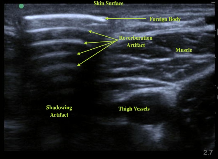
Ultrasound is especially useful in real-time to facilitate foreign body removal. Once located the object should be scanned in two planes to determine its size and proximity to surrounding structures. In this case, while scanning providers identified vessels in the thigh in close proximity to the object so elected not to remove it in the department. This is extremely valuable information that took only minutes to obtain at the bedside.
Date: June 2012